Monday, June 21, 2021

Culture Media PART 3 - Classification Based on Chemical Constituents Part II - DR C R Meera


Based on the chemical constituents used for the preparation, media can be classified into simple media, complex media, defined or synthetic media and semi-defined or semi-synthetic media.

1.    Simple media

Simple media include the basal media or general purpose media like peptone water, nutrient broth and nutrient agar which are very commonly used in Microbiology laboratory for cultivation of microorganisms. Peptone media is prepared using 1% peptone in 0.5% NaCl in water (pH 7.2±0.2).  Components of nutrient broth includes peptone, yeast/ beef extract, sodium chloride and distilled water (pH- 7.2 ± 0.2). When 2% agar is added, it is called nutrient agar. Semi-solid media which is prepared using same components as that of nutrient agar, but with a different concentration of agar, is also considered as a simple media. In semi-solid agar, 0.2 to 0.5% agar is used.  

2.    Complex media

Complex media contain added ingredients for special purposes. It may be for bringing out certain special characteristics of the organisms or for providing special nutrients required for the growth of fastidious organisms. Fastidious organisms are those which are more exacting in their nutritional requirements. Most of the media other than basal media, used in microbiology laboratory are coming under complex media. Eg:  chocolate agar, Blood agar, MacConkey agar, Robertson’s cooked meat (RCM) medium, Lowenstein–Jensen (LJ) medium (Cultivation and isolation of Mycobacterium), etc. Complex media is also called rich media and typically have a mixture of many different organic compounds including glucose as carbon source, a source of amino acids and nitrogen (e.g., beef and yeast extract), various salts, growth factors, vitamins etc. and water needed for bacterial growth. This is an undefined medium, because the amino acid source contains a variety of compounds of which the exact composition is not known. Meat or beef extract is usually made by hydrolysing natural products such as meat with enzymes to release monomers like peptone, tryptone etc from the macromolecules. Another amino acid source yeast extract is made by extracting small molecules from yeast cells. 

3.    Synthetic or Defined media

This media is prepared using pure chemical substances and does not contain any animal, yeast, or plant tissue or their extracts, hence called synthetic media. The exact quantity of all ingredients used in the media is known, hence also called defined media. Synthetic media are used for various special studies such as for studying the metabolic requirements of the organisms. This media consists of a defined carbon and nitrogen source, trace elements (Mn, Mo, Cu, Co, Zn etc.) and vitamins. Glucose or glycerol is often used as carbon source and ammonium salts or nitrates used as inorganic nitrogen sources. Dubos’ medium with Tween 80 used for cultivation of Mycobacterium tuberculosis is an example of this medium. Minimal media (sometimes called Mineral media) is also an example of synthetic media.  Minimal media contain just enough ingredients, generally without the presence of amino acids, to support growth of wild type microorganisms. Minimal media typically consists of  a carbon source (sugar like glucose or less energy source like succinate), various salts with essential element like Mg, N, P, S which help bacteria to synthesize protein and nucleic acids and water.

Supplementary minimal media are minimal media that also contains a single selected agent, usually an amino acid or a sugar. This supplementation allows for the culturing of specific lines of auxotrophic recombinants.

4.    Semi- synthetic or Semi- defined media

Semi-synthetic media will be containing a set of chemical substances of known quantity like the synthetic media, but differ in that at least some natural ingredients of unknown or variable composition are also present.  synthetic medium can be made semi-synthetic by adding a substances such as yeast extract or  beef extract. Eg: Peptone water which is made up of 1% peptone with 0.5% NaCl in water

Video link: https://youtu.be/ubturhQ90Zo


Sunday, June 13, 2021

Culture Media-Part 2-Semisolid Media- Dr Meera C R


Semisolid media have a consistency between Solid and liquid media. It has a soft custard or jelly like appearance.Its components are same as solid media. Only difference is in the concentration of agar used. In solid media 2% agar is used, ie: 2gm in 100 ml of the media. In semisolid media agar concentration used is 0.5% or less. They are used in demonstration of bacterial motility and to grow microaerophilic bacteria.

Bacterial motility is due to the presence of locomotory organelles like flagella. In motility test, bacteria is stabbed using a straight inoculation needle in to the semisolid media and incubated at 370C overnight. After incubation tubes are observed for bacterial growth pattern. Non motile organisms will grow confined to the stab-line, having sharply defined margins and leave the surrounding medium clearly transparent. Motile organisms will move from the line of stab producing diffuse, misty growths that spread throughout the medium rendering it slightly opaque. 


Microaerophiles are bacteria that cannot tolerate atmospheric concentration of oxygen. However they need oxygen for their growth. These groups of bacteria can grow under reduced oxygen (5% to 10%) and increased carbon dioxide (8% to 10%) concentration. Higher amount of oxygen is inhibitory to their growth. Examples: Campylobacter jejuni, Helicobacter pylori, etc. Microaerophiles when inoculated to semisolid media, will move from the surface to the just lower layer where oxygen penetration is only in the required concentration.


Youtube Video: https://www.youtube.com/watch?v=214OubJf1yM&t=1s


Classification of bacteria based on Oxygen requirement- Dr C R Meera

Based on requirement of oxygen for growth, bacteria can be classified into the following groups.

·       Obligate Aerobes: are strict aerobes. Can grow only in the presence of oxygen (21%). Eg. Mycobacterium tuberculosis, Pseudomonas aeroginosa

·       Obligate Anaerobes: are strict anaerobes. Grow only in the complete absence of oxygen. Oxygen presence is toxic and lethal to their growth. Eg: Clostridium perfringens, Clostridium boyulinum.

·       Aerotolerant Anaerobes: Some anaerobic bacteria can tolerate low levels of oxygen and are called nonstringent or aerotolerant anaerobes.  Oxygen is actually not required for their growth but not harmful if present. Eg: Propionibacterium acnes

·       Facultative anaerobes: They can grow well in both aerobic and anaerobic conditions. They do not require oxygen for growth, but may use it for energy production if available.  e.g., Enterobacteriaceae group, Staphylococcus aureus

·       Microaerophiles: Require low levels of oxygen for growth. They can neither tolerate normal levels of oxygen in air nor can live in complete oxygen absence. These groups of bacteria can grow under reduced oxygen (5% to 10%) and increased carbon dioxide (8% to 10%) concentration.  Eg: Campylobacter jejuni, Helicobacter pylori

             Their growth when inoculated into semisolid media is as shown in the diagram.


Wednesday, June 9, 2021

Contributions of Joseph Lister to the field of Microbiology


“Success depends upon attention to detail”- Joseph Lister

Joseph Lister, was a British Surgeon and pioneer in Antiseptic surgery. He is known as the “Father of Modern Surgery”. Lister was born on April 5th, 1827, in England. After finishing his baccalaureate in Medicine in 1852, Lister joined the Royal College of Surgeons, England. In 1859, Lister joined as surgeon in the Glasgow Royal Infirmary, Scotland.

As a surgeon at the Glasgow, Lister observed that he was losing nearly half his amputation cases to sepsis which is due to wound infection.  At that time, people considered ‘miasma’ or ‘bad air’ as the cause of infection. He started experimenting with new ways to prevent wound infection. He started by implementing Florence Nightingale’s most elementary hygienic principles in his hospital. The floors, covered in dirt and mud, were thoroughly cleaned and the windows kept opened for fresh air to circulate. The towels and bed sheets were regularly washed.

He was attracted by Louis Pasteur’s theory that decay and fermentation are results of microbial activity. He concentrated on Pasteur’s theories and experiments and discovered that in the suppuration of wounds as well, micro-organisms play a fundamental role. Lister realised that these microbes had to be destroyed before they enter a wound. This became the basis of his antisepsis doctrine.

Pasteur had recommended filtration, pasteurization and use of antiseptics as the means to remove microorganisms. Motivated by this idea, Lister started trials to find out suitable antiseptic agents that are capable of killing these septic germs and can be safely applied on wounds. At that time, Carbolic acid (phenol) had been used to control putrid sewer stench. Lister experimented with phenol on infected frog legs, and the results were remarkable.

On August 12th, 1865, his first human trial of phenol was carried out on an eleven-year old boy, James Greenlees, brought into the Glasgow Royal Infirmary. Lister treated the wound with phenol and the wound healed without suppuration. Lister recognised the antiseptic properties of phenol and used it during operations as well as for cleaning surgical instruments. Lister started to use carbolic acid to clean hands, clothes and also in operation theatres. Wounds were cleaned and sprayed with phenol before dressing. Lister even invented a carbolic acid spray machine (donkey engine) to produce a mist of carbolic acid in the operating theatre as an attempt to reduce post-operative infections. He noticed that pure phenol is too powerful, and its dilutions up to 1:40 have germicide effect. In his “donkey engine”, he used dilutions up to 1:100 which was effective in air sanitization.

After nine months of experiments, he published his promising data in the International General Medical Journal - Lancet as “Papers on a New Method of Treating Compound Fracture, Abscess, etc., With Observations on the Condition of Suppuration”.  He visited conferences and symposia all over Europe, stating proudly that none of his patients so far have died from sepsis, wound infection or hospital gangrene.


                                                                    “Donkey Engine”

To avoid infection from wound suturing threads, he introduced suturing with catgut instead of the commonly used silver threads. He treated the thread first by prolonged “carbolization”. As an experiment, he sutured deep neck wound in a calf. To his surprise, there was no pus on the suture and also there was no trace of catgut. Catgut suture get naturally degraded by the body's own proteolytic enzymes. Catgut suture threads are derived from strands of purified collagen taken from small intestine of healthy ruminants (cattle, sheep, goats) or from beef tendon.

Honours

·  Chassaignac introduced the use of drainage tubes into surgery. Lister was fascinated by the idea and immediately applied it to the first abscess that came his way. It was not just any abscess; it was located in her Majesty Queen Victoria’s armpit. Using his antiseptic techniques, he treated the abscess so meticulously and he was honoured with “Sir “title.

·       In 1897, Queen Victoria honoured Lister with “Baron” title  

·       First  physician who received the highest noble title of “Lord”

·       In 1899, the “British Institute of Preventive Medicine” was renamed as the “Lister Institute of Preventive Medicine”.

On 10th of February, 1912, Joseph Lister passed away at the age of 85 years.


Tuesday, May 18, 2021

Oxidase Test


Oxidase enzymes play a vital role in the operation of the electron transport system during aerobic respiration.  Cytochrome oxidase catalyses the oxidation of a reduced cytochrome by molecular oxygen (O2), resulting in the formation of H2O or H2O2.  Aerobic bacteria as well as some facultative anaerobes and microaerophiles, exhibit oxidase activity.  The oxidase test aids in differentiation among members of the genera Neisseria and Pseudomonas, which are oxidase positive, and Enterobacteriaceae, which are oxidase negative.

Aim

To distinguish among groups of bacteria on the basis of cytochrome oxidase activity.

Principle

The ability of bacteria to produce cytochrome oxidase can be determined by the addition of the test reagent, p-aminodimethylaniline oxalate, a reagent which serves as an artificial substrate, donating electrons and thereby becoming oxidized to a blackish compound in the presence of the oxidase and free oxygen.  The dark coloration is indicative of cytochrome oxidase production and represents a positive test.  No color change is indicative of the absence of oxidase activity and is a negative test.

Requirements

24 hr nutrient broth cultures of species Escherichia coli, Proteus, Pseudomonas, Staphylococcus, Streptococcus, and Bacillus.

Oxidase strips containing p-aminodimethylaniline oxalate, Bunsen burner, glass rod, glass marking pencil etc.

Procedure

  1. Remove oxidase strips from the container and place in a petridish.
  2. With the help of a clean glass rod / plastic loop or platinum wire pick a colony from 24 hrs growth of      the test organism and rub over the filter paper.  Do not use nichrome wire.
  3. Observe the color change within 5-10 sec. 

Observations

Pseudomonas sp. produced an intense deep blue color within 5-10 sec indicating a positive result.  Escherichia coli, Proteus, Staphylococcus, Streptococcus, and Bacillus sp. produced no change in color.

Result

Pseudomonas sp. is oxidase positive whereas Escherichia coli, Proteus, Staphylococcus, Streptococcus, and Bacillus sp. are oxidase negative.

(Image courtesy: orbitbiotech.com)


Thursday, May 13, 2021

Animal Cell Culture as a Substitute for Animal Experiments- Dr C R Meera


Animals are extensively used for research purposes. They are mainly used for efficacy testing and toxicological screening of drugs for different infectious and non-infectious diseases and for screening of carcinogens. Animals are also sacrificed for medical and surgical experiments. They are also widely used for production of vaccines, antibiotics etc. The number of animals used for experiments have increased considerably with advancement in medical technology & research. Millions of experimental animals are exploited or sacrificed all over the world every year.  Various animals are used for this purpose which include rodents (mice, rats, hamsters, rabbits, guinea pigs), fishes (zebra fish, trout), birds (mainly chicken), amphibians (xenopus frogs), primates, dogs, cats etc. Many animals die during experiment and others are sacrificed at the end of experiments. The pain, distress and death experienced by the animals during scientific experiments have been a debating issue for a long time. Various acts and laws have been passed to bring the control over unethical use of animals and minimize the pain to animals during experimentation. Regulations put forward by the Animal Ethics Committee have to be strictly followed for conducting animal experiments.


Animal cell culture, growing cells in dishes is an excellent alternate for animal experiments. It has many advantages over animal experiments.

  1. Cost and space required are more in animal experiments. Cost of animal experiments are high as breeding, maintenance and care of animals are to be done according to Government rules. Animals have to be maintained in specific breeding centers occupying large areas associated with the research centers. Cell culture is less expensive compared to animal experiments and cells can be grown in small containers. Space consumed for cell culture lab is also very less or even we can work with cell lines on a bench top.
  2. Animal experiments are difficult to perform and require skilled/trained manpower is required to handle the experiments. Cell culture techniques are easy to perform and less manpower required.
  3.       In animal experiments, the number of animals required for a single experiment is many. But in cell culture, many experiments can be done using one animal by dividing the tissue into different experimental groups. Thus multiples of the test can be easily performed.  

    E.g.: Almost all cosmetics, drugs and chemicals are tested for their toxicity and efficacy before commercialization. For example, irritancy test. To check the irritancy of cosmetics/chemicals previously Draize test was used, which requires animals (mainly rabbit). It is very painful and every time a new animal is used. The procedure involves applying 0.5 mL or 0.5 g of a test substance to the eye or skin of a restrained, conscious animal, and then leaving it for set amount of time before rinsing it out and recording its effects. The animals are observed for up to 14 days for signs of erythema and edema in the skin test, and redness, swelling, discharge, ulceration, hemorrhaging, cloudiness, or blindness in the tested eye.  Cell culture replaced it with Corneal organ culture. The bovine cornea is cultured up to three weeks in laboratory and various analytical methods are used to evaluate the toxicological effect of test chemical irritancy in vitro.              Live animals and embryos are used to study effects of some compounds on embryo development. In vitro embryonic stem cell culture test helps to reduce the number of live embryo used and the compounds which are toxic toward developing. 

  4. Another advantage of cell culture is time. Animals experiments have time consuming lengthy protocols and takes considerable time. It usually takes a whole day to do one experiment. Organ or tissue culture experiments are of shorter duration compared animal experiments and done with multiple samples at one time.

  5. .   Tissue culture is having controlled physiochemical environment (pH, temperature, osmotic pressure, O2, CO2 tension etc.) as well as physiological conditions within the system which cannot be controlled in animal experiments. Hence results of tissue culture are more reliable than animal experiments.

  6.     Cell cultures are more suitable for screening the efficacy or toxicity of drugs, carcinogens and chemicals.  In cell culture, the compound to be screened is cultured with isolated organ, tissue or cells. In such systems, it is easy to understand the response of cell line towards the drug. In the whole animal, it is not easy to determine the specific effect of a particular drug. Because, it is possible that known or unknown endogenous substances in the animal body may be interacting with the exogenous material being tested.

  7.         Also less reagents are required in cell culture which are readily available to target cells. In animal experiments, 90% injected chemical is lost by excretion and distribution to tissues other than target cells. In cell culture, cells are exposed directly to the reagent at a lower and defined concentration and with direct access to the target cells.

  8.     In vitro cell culture is a good way to screen the compounds at preliminary stages. For example, use of the human hepatocyte culture gives the information about how a drug would be metabolized and eliminated from the body. Such experiments help to eliminate unsuitable compounds from further studies and thus minimizes the use of animals in further experiments.

  9.       Tissue samples collected from animals for experiments are invariably heterogeneous. But cultured cell lines, after one or two serial passages assume a homogeneous or uniform constitution which reduces the need for statistical analysis of variance. 

Wednesday, May 5, 2021

Asexual Reproduction in Fungi- Dr C R Meera

 Asexual Reproduction in fungi


Fungi reproduce by both sexual and asexual means. 

  • Yeasts which are unicellular fungi reproduce by budding which is an asexual process. Eg: Cryptococcus neoformans. Some yeasts like Schizosaccharomyces pombe reproduce by fission instead of budding and thereby two identically sized daughter cells are produced. Schizosaccharomyces pombe is a facultative sexual organism and undergoes sexual reproduction under high-stress conditions such as nutrient starvation. In this condition yeast haploid cells will die. However, diploid cells which are formed by conjugation undergo sporulation, entering sexual reproduction (meiosis) and producing a variety of haploid spores, which can go on to mate (conjugate), reforming the diploid.

  • Yeast like fungi reproduce asexually by  budding and fission.

  • Molds, the filamentous fungi reproduce by both sexual and asexual methods.

  • Fungi imperfecti, also called deuteromycetes or hyphomycetes, is a provisional group of fungi whose sexual phases have not been identified.  


  1. Asexual Reproduction

Asexual reproduction is also known as somatic or vegetative reproduction. In asexual reproduction there is no union of  nuclei, sex cells or sex organs of two different cells. Daughter cells arise from the single parental cells.  Asexual methods of reproduction include

  1. Fission

  2. Budding

  3. Fragmentation

  4. Spore formation (Asexual spores)


  1. Fission

In fission, the parent cell elongates and divides transversely into two daughter cells of identical size. First, the nucleus divides which is known as Karyokinesis, followed by the division of the cytoplasm and wall formation, known as cytokinesis. In this process, initially the replicated DNA molecule attaches each copy to a different part of the cell membrane. The original and replicated genomes are pulled apart and separated as the cell elongation takes place prior to cell division. All daughter cells produced by fission are genetically identical, meaning that they have the same genetic material. Unlike the processes of mitosis and meiosis used by eukaryotic cells, binary fission takes place without the formation of a spindle apparatus on the cell. Like in mitosis (and unlike in meiosis), the parental identity is preserved.


  1. Budding


Budding is the process in which somatic cells produce a small bud like out growth which develops into a new individual. In this method daughter cells pinch or bud from the parental cell and are smaller than the mother cell. Under optimal conditions, yeast cells divide as rapidly as once every 90 min through a process of budding.


  1. Fragmentation


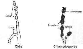
In fragmentation, disjoining of hyphal cells take place and each cell develops into a new organism. Disjoined single cells act as the spores and germinate to produce hyphae and mycelium. Thus from each fragmented cell, a new organism arises. Spores produced by fragmentation are called Oidia or Arthrospores or Thallospores (Eg: Erysiphe).


  1. Spore formation (Asexual spores)

Asexual spores are produced from single parental cell and their function is to disseminate the species. Fungi that produce more than one type of spores are called pleomorphic or polymorphic fungi as they can exist in different morphological forms. 

Spores are produced at the tip of special hyphae called Sporophores. Name of this special hyphae changes according to the type of spore produced on it.

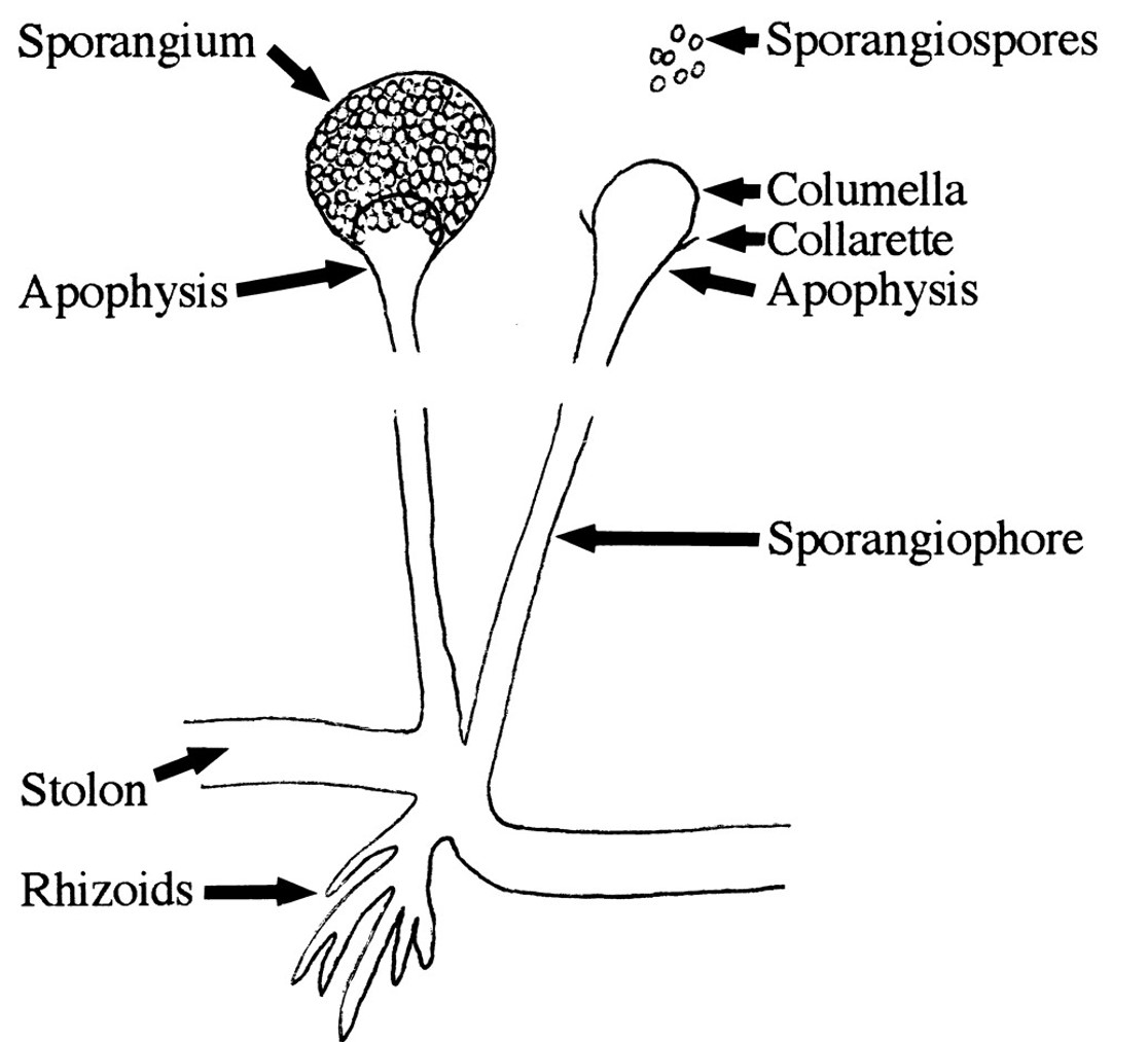
For Eg: Sporophores that produce Sporangiospores are called Sporangiophores.

    Sporophores that produce Conidiospores are called Conidiophores.


Sporophores bear spores either packed inside special sac-like structures called Sporangium (Pl. Sporangia) or at the terminal ends.  If spores are produced inside the sporangia, they are called endogenous spores and if the spores develop exogenously on the terminal ends of sporophores, they are called the exogenous spores. The sporangia may be terminal or intercalary in their position.


Different types of asexual spores are produced by fungi. They include:

a)Sporangiospores

b)Conidiospores or conidia

c)Oidia (Arthrospores/Thallospores)

d)Chlamydospores

e)Blastospores


a)Sporangiospores   

Sporangiospores are single celled spores produced inside the special sac-like structures called Sporangium (Sporangia Pl.). Sporangia are produced at the tip of  special hyphae called Sporangiophores. A sterile dome-like structure at the tip of a sporangiophore or within a sporangium is called Columella. A funnel-shaped swelling of a sporangiophore, immediately below the columella, can be seen in some fungi and are called Apophysis.

Sporangiospores can be motile or nonmotile. Non-motile sporangiospores are called Aplanospores. Motile ones are called Zoospores and their motility is due to the presence of flagella.


b)Conidiospores or conidia (Conidium)

Conidia are exogenous spores formed at the tip or side of hyphae. They can exist in two forms such as Microconidia and Macroconodia. Microconidia are small single celled conidia whereas Macroconidia are large multi celled spores.


C & d)Oidia/ Oidium (Arthrospores/Thallospores) and Chlamydospores


Both Oidia and Chlamydospores are produced by the disjoining or fragmentation of vegetative hyphae. In oidia formation, hyphal cells simply disjoin from the apical regions and each fragmented cell acts as spore, giving rise to new fungi. In the case of Chlamydospores, cells become enveloped by a thick wall before hyphal fragmentation. These thick walled, single celled Chlamydospores are highly resistant to the adverse conditions and can be terminal or intercalary.

e) Blastospores

Blastospores are spores formed by budding. It is also known as a blastoconidium (pl. blastoconidia). An example of a fungus that forms blastospores is Candida albicans.


Other means of asexual reproduction in fungi include formation of Sclerotia and Rhizomorphs. These asexual methods are usually used to overcome unfavourable environmental conditions. 

The sclerotia are resistant and perennating bodies. Each sclerotium is a cushion-like structure of compact mycelium. They survive for many years.  They give rise to new mycelia on the approach of favourable conditions.


Rhizomorphs are rope-like modified mycelium, also resistant to unfavourable conditions and give rise to new mycelia even after several years on the approach of favourable conditions.


Youtube Link: https://www.youtube.com/watch?v=yX7Ba5VCI9w


Complement System: Activation & Functions

1. INTRODUCTION Complement (C): A system of factors that occur in normal serum. Activated by Antigen–Antibody (An–Ab) interaction. Subsequen...