Showing posts with label Molecular Biology. Show all posts

Sunday, January 8, 2023

DNA as Genetic Material-Experimental Proof Part III- The Blender Experiments- Dr. C R Meera


DNA as Genetic Material-Experimental Proof

Part III- The Blender Experiments- Dr. C R Meera

An elegant confirmation of genetic nature of DNA came from the experiments with E.coli phage T2 by Hershey and Chase.

·        This experiment is called Blender experiment as “Kitchen Blender” was used as the major apparatus.

·        Done by Alfred Hershey and Martha Chase, who demonstrated that DNA injected by phage particle into bacterium contains all information required to synthesize progeny phage particles.

·        T2 phage used in the experiment consisted of DNA encased in a protein shell.

·        Radioactive phosphate and sulfur were used to mark the phage.

·        DNA is the only phosphorous-containing particle in the phage. So radioactive phosphate would be incorporated in phage DNA during multiplication in a media containing radioactive phosphate .

·        Proteins of the shell which contain the amino acids methionine and cysteine, have only sulfur atoms in it. So protein shells would take up the radioactive sulfur during multiplication in a media containing radioactive sulfur.

·        In this experiment, Phage with radioactive DNA was used which was prepared by growing phages in a nutrient medium in which radioactive phosphate          (32PO43-) is the sole source of phosphorus.

·        Phage with radioactive capsid- was also used which was prepared by growing phages in a medium containing radioactive sulfur (35SO42-) as the sole source of sulfur.

·        These two kinds of radiolabeled phages were used in the infection of E.coli so that phage DNA and capsid can be located by the radioactivity.

·        T2 phage has a long tail with which it attaches to the host bacterium, E.coli. Hershey and Chase showed that the attached phage can be separated from the bacteria by violent agitation using the kitchen blender.

·        They conducted two experiments known as 35S and 32P Experiments.


                                   Fig 1. 35S and 32P Experiments by Hershey and Chase                                         
  (Image Courtesy:www.mun.ca)


35S Experiment

·         Phage with radioactive capsid was also used in this experiment which was prepared by growing phages in a medium containing radioactive sulfur.

·         S labeled phages were allowed to adsorb to bacteria for a few minutes.

·         Phage-attached bacteria were separated from unattached phages by centrifugation of the mixture and the pellet formed is the phage-bacterium complex.

·         This complex was resuspended in a liquid and blended and the suspension was again centrifuged.  

·         Now, the pellet received is bacteria and the supernatant was also collected.

·         80% of the radioactivity or  35S was in the supernatant and 20% was in the pellet. This experiment thus showed that capsid remains outside the bacteria and not acting as the genetic material.

·         Many years later, it was found that 20% radioactivity in the pellet was due to the tail segment of the phage being too tightly attached to the bacterial cell that were not removed by blending.

32P Experiment

·         Phage with radioactive DNA was used in this experiment which was prepared by growing phages in a nutrient medium containing radioactive phosphate. The experiment was performed same as above.

·         A very different result with 70% of radioactivity or 32P was found in the pellet (ie; associated with the bacteria) and 30% radioactivity in the supernatant.

·         30% radioactivity in the supernatant may be due to the breakage of bacteria during the blending process or due to the defective phage particles that could not inject their DNA into the bacterial host.  

·         This experiment proved that the DNA is getting into the bacterial cell and hence acting as the genetic material.

·         Confirmation Experiment: Pellet was resuspended in the growth medium and re-incubated. It was capable of phage production, indicating that the genetic message for phage progeny production had been introduced by phage DNA, not by phage protein. Thus, DNA is the genetic material.

·         Later, a series of experiments called “transfer experiments” analyzed the progeny for 35S and  32P.

·         35S was not found in the progeny.

·         Half of  32P injected was found in the progeny.

·         Interpretation: Transfer of only half of 32P in progeny is because that DNA is selected at random for packing into protein coats.


DNA as Genetic Material-Experimental Proof Part II- The Chemical Experiments- Dr. C R Meera

 DNA as Genetic Material-Experimental Proof                                                         Part II- The Chemical Experiments- Dr. C R Meera                                                       

Chemical experiments to prove DNA as genetic material was put forward by Chargaff & collegues in 1940s.

In 1930s, existed the Tetranucleotide hypothesis by Phoebus Levene suggested that DNA is composed of repeating sequences of four nucleotides. The hypothesis suggested that DNA contained equimolar quantities of Adenine, Thymine, Cytosine, and Guanine. There were two reasons behind the development of such a hypothesis.                                                                     1.           Techniques used to separate bases did not resolve them very well, so quantitative analysis was not perfect.

2.           DNA analyzed was isolated from mainly eukaryotes in which four bases are equimolar or from bacteria in which bases were almost equimolar.

The most important clue to the chemical structure of DNA came from the work of Erwin Chargaff and colleagues in the 1940s.  Using the DNA of a wide variety of organisms, Chargaff applied new separation and analytical techniques and showed that molar concentrations of bases could vary widely. Thus, DNA could have variable composition, a primary requirement for genetic material. They found that 4 nucleotide bases of DNA occur in different ratios in the DNA of different organisms. They also found that the amounts of certain bases are closely related. Data collected from so many different species lead Chargaff to the following conclusions known as “Chargaff’s Rules”.

1.           The base composition of DNA generally varies from one species to another.

2.           DNA specimens from different tissues of the same species have the same base composition

3.           The base composition of DNA in a given species does not change with organism’s age, nutritional state, or changing environment

4.           In all cellular DNAs,   regardless of the species, the number of adenosine residues is equal to the number of thymidine residues (A=T) and the number of guanosine residues is equal to cytidine residues (C=G). Thus, the sum of purine residues = sum of pyrimidine residues.

ie;   (A + G)  = (C + T)

Upon publication of Chargaff’s results, Levene’s tetranucleotide hypothesis quietly died and the idea of DNA as the genetic material began to catch on.

Shortly after, researchers in several laboratories found that, for a wide variety of organisms, somatic cells have twice the DNA content than germ cells, a characteristic expected of the genetic material.

Thus, objections to the work of Avery, McLeod, and McCarty were no longer heard and the hereditary nature of DNA rapidly became the acceptable idea.


DNA as Genetic Material-Experimental Proof-Part I-Dr C R Meera


DNA as Genetic Material-Experimental Proof

Part I- The Transformation Experiments- Dr. C R Meera

Three simple experiments, done with great care, identified DNA as genetic material. They included:

1.    The transformation experiments (Griffith/Avery, McLeod & McCarty)

2.    The chemical experiments (Chargaff)

3.    The Blender experiments (Hershey & Chase)

I.     The Transformation Experiments

The first experimental proof for DNA as genetic material was by Fred Griffith in 1928. He was studying the human bacteria causing pneumonia, Streptococcus pneumoniae or Pneumococcus. Its virulence is attributed to its polysaccharide capsule that protects it from body defense. On Nutrient agar media, the organisms produced colonies with smooth edges due to the presence of capsules and are called “S colonies”. When these organisms were injected into mice, it caused the death of the animal. This means, S colonies are lethal.

Griffith could isolate mutants of the organisms which were producing rough-edged colonies and named them “R bacteria”. They were non-encapsulated and non-lethal when injected into the mice.

He further experimented with “heat-killed S” colonies which were again non-lethal as “R colonies”. A significant observation made by Griffith was that when he injected the mixture of  “R live” and “heat-killed S”, it resulted in the death of the mice. He also isolated the bacteria from the dead mice and surprisingly it was found to be “S live”. He thought that the “R live” was replaced or transformed into “S live” forms within the mice.  

After several years, it was found that mice are not necessary for this transformation. The same experiments were carried out in In vitro models.  “R live” and “heat-killed S” cells were grown together in culture media and “S live” cells could be isolated from the same. And it was concluded that “R cells” restored the viability of “S cells”. But this idea was eliminated later due to another experiment in which “R cells” and “cell extract of heat-killed S cells”(extract was prepared from broken S cells and freed from both intact cells and capsular polysaccharides) produced “S live” strain. This experiment concluded the “cell extract” as the “transforming principle” nature of which was unknown at that time. 


Fig.2. Experiment with live “R cells” and “cell-free extract of lysed S cells”

The next development occurred some 15 years later in 1944, when Oswald Avery, Colin McLeod, and Maclyn McCarty partially purified the transforming principle from cell extract and demonstrated that it was DNA.

However biochemical investigation of DNA had begun in 1868 with Freidrich Miescher.

Ø  Miescher isolated a phosphorous-containing substance from nuclei of pus cells or leukocytes in the discarded surgical bandages. He named it “nuclein”. Nuclein consisted of acidic and basic portions. The acidic portion is now known as DNA and the basic portion is protein.

Ø  Later, Miescher discovered the same “acidic” substance in the heads of the sperm cells of salmon. However, he could partially purify nuclein and studied its properties. However, its primary covalent structure was not known till 1940s.

The first direct evidence for DNA as the bearer of genetic information came in 1944 by the experiments of Oswald Avery, Colin McLeod, and Maclyn McCarty. They extracted DNA from heat killed S strain (virulent), protein was removed as much as possible. It was then mixed with non-virulent R strain. DNA gained entrance into the R cells by the process of transformation (transformation is defined as the genetic alteration of a cell which is caused by the direct uptake and incorporation of exogenous genetic material from its surroundings through the cell membrane). In this experiment “non-virulent R strain” was permanently transformed into the “virulent S strain”.   The procedure for purifying DNA at that time was not perfect and contained many impurities. Hence, necessary evidence for transformation by DNA was given by Avery, McLeod, and McCarty using the following procedures.

1.    Chemical analysis of transforming principle- Chemical analysis revealed the major component as deoxyribose containing nucleic acids

2.    Physical measurement – The sample contained highly viscous substance with physical properties of DNA

3.    Enzyme reactions- Transforming principle was treated first with proteolytic enzymes like trypsin, chymotrypsin, or both.

Also treated with ribonuclease (RNA depolymerizing enzyme). Transforming activity was not lost when the treated transforming principle was mixed with the R strain, indicating that neither proteins nor RNA is the active principle.

Treatment with DNase (catalyze the hydrolytic cleavage of phosphodiester linkages in the DNA backbone and degrade DNA) inactivated the transforming principle which was a shred of clear evidence for DNA as genetic material.

Through these procedures, Avery, McLeod, and McCarty concluded that the transforming principle they isolated is DNA.

But the scientific community of that time was not ready to accept their conclusions. Because, whatever the genetic material was, it was expected to be a substance capable of the enormous variation in order to contain information carried by the huge number of genes. At that time, DNA was known as a tetranucleotide only (tetranucleotide hypothesis), so could not be considered the sole material of genetic information.  

Early experiments suggested that 4 bases (adenine (A), cytosine (C), guanine (G), and thymine (T)) occur in equal ratios in nucleotides. After discovering the location of  nucleic acids on chromosomes, the tetranucleotide hypothesis was put forward by Phoebus Levene in 1929. Ribose sugar and Deoxy ribose sugar were discovered by him in 1909 and 1929, respectively.    In the tetranucleotide hypothesis, Levene suggested that nucleic acids are repeating tetramers.  In other words, DNA is composed of repeating sequences of four nucleotides. He called the Sugar-Base-Phosphate unit a nucleotide. Also said that the simplicity of this structure of DNA was too uniform to contribute to a complex genetic variation. Hence, DNA could not be the genetic material.  Thereafter, attention focused on proteins as the probable hereditary substance.

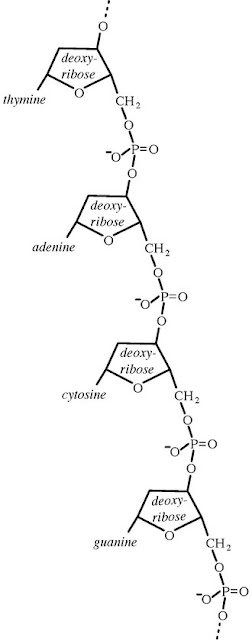
Fig 3. Tetranucleotide hypothesis: Repeating tetranucleotide unit

(Image courtesy: en.wikipedia.org)


Monday, May 25, 2020

Eukaryotic Chromosome Organization


1. Eukaryotic and prokaryotic cells differ in a number of aspects. In Greek, Eu means true, Pro means before and karyon means nucleus. Main difference is in their nucleus. Presence of a well-defined nucleus is the main feature that distinguishes the eukaryotic cell from a prokaryotic one. Nucleus serves as the cell’s control center and genetic information is packed up there. DNA replication, transcription, RNA processing etc take place in the nucleus, and only final stage of translation occurs in cytoplasm.      

Eukaryotic nucleus has nuclear membrane, chromatin and nucleolus which is the most prominent substructure in nucleus. Nucleolus are cell’s ribosome factories.  Chromatin, RNA and nuclear proteins move freely in aqueous solution in nucleus.  

·     In prokaryotes, usually a single chromosome is present per cell in the form of covalently closed circular DNA. They also contain extra chromosomal DNA called plasmids. But in eukaryotes, several chromosomes are present per cell (2 to many). Here also extra chromosomal DNA is found in mitochondria and chloroplast. Amount of chromosome in eukaryotic cells is much more than a prokaryotic cell. A human cell contains DNA more than a 1000 times than E coli. In E coli 4x10⁶ base pairs (bp) are seen in single cell whereas in human cell 5.5x10⁹ bp of DNA is present in its diploid nucleus. Each eukaryotic species has a characteristic number of chromosomes.
·     3. C Value- For eg: In humans 46 chromosomes (diploid set) are seen, ie., 23 pairs of chromosomes. Half or haploid set is derived from the egg and half from sperm cell. Eukaryotic chromosomes exist as a single linear unbroken double stranded DNA (DS DNA) molecule running throughout the length of chromosome, coupled with double amount of protein. Chromosomes are not always distinct in a cell. They are well defined structures at the time of cell division. At other times, chromosomes are not well organized. Total amount of DNA in a haploid genome of a species is called the “Species C value”. In other words, C value is DNA contained within a haploid nucleus (e.g. a gamete) or half the amount of DNA in a diploid somatic cell of a eukaryotic organism. C value of human beings is 3.4 x 10⁶ kb. 
·                        ·     5. Chromosome organization in Eukaryotic cells- In eukaryotes, cell cycle has 4 phases viz. G1, S, G2 and M phase. G1 phase is also called the first gap phase. In this phase cells grow in size, copies organelles and makes the molecular building blocks needed in later steps. During S phase, the cell synthesizes a complete copy of the DNA in its nucleus. During the second gap phase G2, the cell grows more, makes proteins and organelles, and begins to reorganize its contents in preparation for mitosis. G1, S and G2 phases together are known as interphase.  In other words, interphase is the time between two mitotic divisions. As mitosis begins, G2 phase stops. Mitosis has 4 phases called prophase, metaphase, anaphase and telophase. Mitosis is followed by Cytokinesis, the process of the parent cell division into 2 daughter cells. In G1 phase chromosome is single. During S phase, chromosome duplicate to produce two sister chromatids held together by centromere. Chromosome continues in the same structure in G2 phase. Centromeres separate and sister chromatids become daughter chromosomes in M phase. 

·                   6. In G1 phase chromosome exist as one linear DS DNA molecule. This DNA is usually complexed with double amount of proteins. In S phase, sister chromatids have one linear DS DNA running the length of each chromatid. DNA is least organized in G1 phase and most organized just before cell division. DNA is usually coupled with proteins. DNA and proteins together form the structure called chromatins.  Chromatins are stainable material in cell nucleus and amount of protein is twice the amount of DNA. Basic structure of chromatin in all eukaryotes is identical.
·     7. Chromatin structure- In chromatin, two types of proteins are associated with the DNA called histones and non-histones. Both proteins play a crucial role in the physical structure of the chromosome. Histones are the most abundant proteins. They are small basic proteins with overall positive charge. Large amount of arginine and lysine residues are responsible for the net positive charge and this property helps histones to bind with the negatively charged DNA.  5 types of histones are found in eukaryotes namely H1, H2A, H2B, H3 and H4. In chromatin amount of histone is equal to amount of DNA. Remaining protein part of chromatin is made up of non-histones. Amount and proportion of histone relative to DNA is constant from cell to cell in eukaryotes.
·     8. Among these 5 types of histones, amino acid sequences of H2A, H2B, H3 & H4 are very similar & highly conserved between even distantly related species. H3 & H4 are among the most conserved proteins.  H2A and H2B show slight species specific variation in sequence. This evolutionary conservation shows that histones play some basic role in eukaryotes in the organization of chromosomes. DNA of chromosomes in a single human cell measures >2m (6.5 feet). Human DNA is 700 times greater than E coli. Several levels of packing is necessary to reduce it into cm or even mm to nm and to pack into the nucleus of the cell. Histones play a very crucial role in this chromatin packing.
·     9. Non- histones- These are less abundant than histones. All other proteins associated with DNA apart from histones are called non- histone proteins. These include the proteins that bind during DNA replication, repair, transcription, gene regulation, recombination etc. They are mainly negatively charged. So will bind usually with the positively charged histones. Many different non-histones are present in each eukaryotic cell. Amount and types of histones vary from organism to organism. They also differ markedly in number & type from cell to cell in same organism. Even they differ at times in the same cell.
·     10. Levels of packing in Chromatin - Chromatin structure differs in different stages of the life cycle. It is least organised in the G1 phase and highly condensed at the metaphase, the phase just before cell division. The least compact structure is called 10 nm chromatin fibre. It is morphologically similar to beads on a string. Diameter of bead region is 10 nm and it forms the core region of nucleosomes which are the basic structural units of chromatin. 

·     11. Bead like region is wound around by the string of DNA. This structure is called nucleosome and its diameter is 11nm. Bead like core region consists of 8 histone proteins – 2 each of H2A, H2B, H3 & H4. Also called core histones. This core region is surrounded by DNA of 147bp length wound around by 1.65 times. This arrangement makes the linear DNA compact by a factor of about 6. Thus nucleosome formation is considered as the 1st level of chromatin packing. In other words, 10nm chromatin fibre formation is the first level of chromatin packing. Strands of DNA between nucleosomes are called “linker DNA”. Means two nucleosomes are connected by linker DNA strands. Amount of linker DNA not constant within & among organisms. In humans, linker DNA range between 38-53 bp, thus total amount of DNA per nucleosome is between 185-200 bp. 

·     12. Second level of chromatin condensation is by the formation of “30 nm chromatin fibre” or “Solenoid fibre” which is brought about by H1 Histones.  H1 are larger than other histones. H1 binds to linker DNA at one end of nucleosome and to middle of DNA segment around histone core, making the 10 nm chromatin fibre more compact.   A nucleosome core plus H1 is called Chromatosome. This condensed DNA spiral helically & becomes regular pattern with 6 nucleosome per turn. Now the diameter of DNA is  30 nm and is called “30 nm chromatin fibre” or “Solenoid fibre”.  Solenoid fibre is seen during interphase of cell cycle. This structure brings the packing ratio of DNA to ~40.

 · 13. Next level of chromosome condensation is by the formation of Chromosome Scaffold. However, the exact mechanism behind it is not clearly understood. Non-histone proteins are mainly involved in Scaffold formation. Non histone proteins bind to 30 nm chromatin fibre to form chromatin loops of 30-90 kb long DNA.  180-300 nucleosome will be usually present per loop. In humans around 2000 such looped domains are present. Stretches of DNA that bind to non-histone proteins to form the loop are called Scaffold associated Regions (SAR).  These chromatin loops attach spirally around the Chromosome Scaffold which is again made up of non-histone proteins and form “Rosettes of chromatin loops”. Around 15 loops are present per turn. This makes the chromosomes now 10,000 times shorter & 400 times thicker than naked DNA. 

·     14. Naked DS DNA is 2nm in its diameter but around 2m long. 10-20 µm is the size of a typical eukaryotic cell and its nucleus is even smaller, having a size ranging between 2-10 µm. So, in order to accommodate the very long DNA into this very small nucleus of eukaryotic cell, it has to undergo different levels of packing.  When it undergoes first level of folding into nucleosome or Bead-on-string pattern, the diameter is 11 nm. Means DNA is getting thicker but even shorter. When chromatin undergoes second level of folding into solenoid fibre, its diameter becomes 30 nm. Further folding in to scaffold loop, makes it to a dimension of   300nm in diameter. Further spiralling of loop around chromosome scaffold makes DNA 700nm in diameter. Entire condensed chromosome is 1400 nm in diameter as consists of two sister chromatids. Such 46 chromosomes are packed in the nucleus of each eukaryotic cell (somatic cells).
·     15. To Summarise, in first level of chromatin packing, DS DNA is wound around histones to form nucleosomes and 10 nm chromatin fibre is produced. H1 histones cause second level of condensation of chromatin into 30 nm chromatin fibre or Solenoid fibre. Third level of packing is brought about by non-histone proteins and Scaffold loops are formed which helically get arranged around the chromosome scaffold.  Thus DNA in chromosome becomes 10,000 times shorter and 400 times thicker and accommodated in the nucleus of eukaryotic cell.

Watch My Video on- Eukaryotic Chromosome Organization


General Methods of Classification-Dr C R Meera

Ø     Goals of Classification A classification system should have two qualities. a.               Stability b.              Predic...