Monday, July 10, 2023

General Methods of Classification-Dr C R Meera


Ø    Goals of Classification

A classification system should have two qualities.

a.              Stability

b.             Predictability

Stability- Frequent changes in the classification system can cause confusion. Therefore, it is important to strive for the development of a classification system that requires only minor adjustments when new information becomes available. This will help minimize confusion and ensure stability in the classification system.

Predictability- By knowing the characteristics of one member of a taxonomic group, it should be possible to assume that the other members of the same group probably have similar characteristics.

Predictability is an important characteristic of a classification system. It refers to the ability to anticipate and understand how the system categorizes and organizes information. A predictable classification system follows consistent rules and criteria, allowing users to have a clear understanding of how entities are classified and how they relate to each other.

Ø    General Methods of Classification

1.             Intuitive method

2.             Phenetic or Phenotypic classification

3.             Phylogenetic or Phyletic classification

4.             Genotypic or Genetic classification

5.             Numerical taxonomy or Adansonian classification

 1.             Intuitive method

         In this method, a microbiologist who has been studying the properties of the organisms for several years may decide that the organisms under study represent one or more species or genera. The drawback of this system is that the characters considered important by one person may not be so important to others. So different taxonomists may arrive at very different groupings. This is a primitive method that is not in use now.  However, some schemes based on intuitive methods have proved to be useful.

2.             Phenotypic classification

      For a very long time, microbial taxonomists relied on this system. This classification system is based on the mutual similarity in the phenotypic or morphological characteristics of the organisms. This system succeeded in bringing order to biological diversity. To some extent, this system also clarified the function associated with morphological structures. For eg: Flagella and motility are associated in most organisms. So, it could be assumed that flagella are involved in at least some types of motility.

Phenetic studies can also reveal evolutionary relationships. However, this system is not dependent on phylogenetic analysis. The phenetic system is based purely on morphological characters. This system will compare as many traits as possible, giving equal weightage to all traits studied. The best phenetic classification is the one constructed by comparing as many attributes/ morphological characters as possible. Organisms sharing many characteristics (>70%) are grouped into a single taxon.

3.             Phylogenetic or Phyletic classification

      With the publication of Darwin’s “On the Origin of Species’ in 1859, biologists began to develop phylogenetic or phyletic classification system. This classification system is based on the evolutionary relationships of organisms. The term “phylogeny” means ‘evolutionary development of a species’. In Greek, ‘phylon’ means ‘tribe or race’ and ‘genesis’ means ‘origin or generation.’ However, for much of the 20th century, microbiologists could not effectively employ phylogenetic classification systems mainly due to the lack of good fossil records. When Carl Woese and George E Fox proposed the use of rRNA nucleotide sequences to assess evolutionary relationships among 
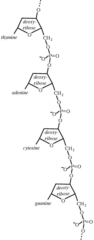
microorganisms, the doors opened for phylogenetic classification systems. 16S ribosomal RNA (or 16S rRNA) is the RNA component of the 30S subunit of a prokaryotic ribosome (SSU rRNA). It binds to the Shine-Dalgarno sequence and provides most of the SSU structure. The genes coding for it are referred to as 16S rRNA gene and are used in reconstructing phylogenies, due to the slow rates of evolution of this region of the gene. The validity of this approach is widely accepted and over 2 lakh different 16S and 18S rRNA sequences are saved in the International databases-  GenBank and the Ribosomal Database Project (RDP-II).

4.             Genotypic or Genetic classification

     In the genotypic classification system, genetic similarity between the organisms is evaluated. In this method comparison of either the individual gene or whole genome (haploid set of chromosomes in a microorganism) is done. Since 1970, it is widely accepted that if the genome of prokaryotes shows similarity in more than 70%, they could belong to the same species. Microbial genomes could be compared in many ways. The simplest method is DNA Base Composition determination. Other ways include Nucleic acid hybridization, Nucleic acid sequencing, and Genomic fingerprinting (evaluation of genes that evolve more quickly than those that encode rRNA.

5.             Numerical taxonomy or Adansonian classification

     Numerical taxonomy, also known as numerical phenetics or phenetic taxonomy, is a method used in biological classification to group organisms based on their overall similarity with the help of computers. Unlike traditional taxonomy, which relies on morphological characteristics and evolutionary relationships, numerical taxonomy employs quantitative data from various attributes or characteristics of organisms. This approach aims to create classifications based on the overall similarity or dissimilarity of organisms rather than their shared ancestry.

The process of numerical taxonomy involves several steps:

1. Data Collection: Quantitative data is collected from the organisms under study. This data can include various traits such as morphological features, biochemical characteristics, physiological measurements, or genetic markers.

2. Data Standardization: The collected data is standardized to ensure compatibility and comparability across different organisms. This step may involve converting measurements to standardized units or transforming data to eliminate biases or variations.

3. Similarity or Dissimilarity Calculation: A similarity or dissimilarity matrix is constructed based on the standardized data. Different mathematical methods can be used to calculate the degree of similarity or dissimilarity between pairs of organisms. Common methods include the Jaccard coefficient or correlation coefficients.

4. Cluster Analysis: Cluster analysis is performed using the similarity or dissimilarity matrix to group organisms into clusters or taxa.

5. Taxonomic Hierarchy: The clusters obtained from the cluster analysis are organized into a taxonomic hierarchy. This hierarchy can include higher-level groupings, such as classes or orders, as well as lower-level groupings, such as genera or species.

Numerical taxonomy has been widely used in fields such as ecology, microbiology, botany, and zoology. It provides a systematic and quantitative approach to classify organisms based on overall similarities, without relying on subjective interpretations or expert judgments. However, it is important to note that numerical taxonomy does not consider evolutionary relationships explicitly, which can be a limitation in certain contexts where phylogenetic information is crucial for understanding the evolutionary history of organisms.


Sunday, January 8, 2023

DNA as Genetic Material-Experimental Proof Part III- The Blender Experiments- Dr. C R Meera


DNA as Genetic Material-Experimental Proof

Part III- The Blender Experiments- Dr. C R Meera

An elegant confirmation of genetic nature of DNA came from the experiments with E.coli phage T2 by Hershey and Chase.

·        This experiment is called Blender experiment as “Kitchen Blender” was used as the major apparatus.

·        Done by Alfred Hershey and Martha Chase, who demonstrated that DNA injected by phage particle into bacterium contains all information required to synthesize progeny phage particles.

·        T2 phage used in the experiment consisted of DNA encased in a protein shell.

·        Radioactive phosphate and sulfur were used to mark the phage.

·        DNA is the only phosphorous-containing particle in the phage. So radioactive phosphate would be incorporated in phage DNA during multiplication in a media containing radioactive phosphate .

·        Proteins of the shell which contain the amino acids methionine and cysteine, have only sulfur atoms in it. So protein shells would take up the radioactive sulfur during multiplication in a media containing radioactive sulfur.

·        In this experiment, Phage with radioactive DNA was used which was prepared by growing phages in a nutrient medium in which radioactive phosphate          (32PO43-) is the sole source of phosphorus.

·        Phage with radioactive capsid- was also used which was prepared by growing phages in a medium containing radioactive sulfur (35SO42-) as the sole source of sulfur.

·        These two kinds of radiolabeled phages were used in the infection of E.coli so that phage DNA and capsid can be located by the radioactivity.

·        T2 phage has a long tail with which it attaches to the host bacterium, E.coli. Hershey and Chase showed that the attached phage can be separated from the bacteria by violent agitation using the kitchen blender.

·        They conducted two experiments known as 35S and 32P Experiments.


                                   Fig 1. 35S and 32P Experiments by Hershey and Chase                                         
  (Image Courtesy:www.mun.ca)


35S Experiment

·         Phage with radioactive capsid was also used in this experiment which was prepared by growing phages in a medium containing radioactive sulfur.

·         S labeled phages were allowed to adsorb to bacteria for a few minutes.

·         Phage-attached bacteria were separated from unattached phages by centrifugation of the mixture and the pellet formed is the phage-bacterium complex.

·         This complex was resuspended in a liquid and blended and the suspension was again centrifuged.  

·         Now, the pellet received is bacteria and the supernatant was also collected.

·         80% of the radioactivity or  35S was in the supernatant and 20% was in the pellet. This experiment thus showed that capsid remains outside the bacteria and not acting as the genetic material.

·         Many years later, it was found that 20% radioactivity in the pellet was due to the tail segment of the phage being too tightly attached to the bacterial cell that were not removed by blending.

32P Experiment

·         Phage with radioactive DNA was used in this experiment which was prepared by growing phages in a nutrient medium containing radioactive phosphate. The experiment was performed same as above.

·         A very different result with 70% of radioactivity or 32P was found in the pellet (ie; associated with the bacteria) and 30% radioactivity in the supernatant.

·         30% radioactivity in the supernatant may be due to the breakage of bacteria during the blending process or due to the defective phage particles that could not inject their DNA into the bacterial host.  

·         This experiment proved that the DNA is getting into the bacterial cell and hence acting as the genetic material.

·         Confirmation Experiment: Pellet was resuspended in the growth medium and re-incubated. It was capable of phage production, indicating that the genetic message for phage progeny production had been introduced by phage DNA, not by phage protein. Thus, DNA is the genetic material.

·         Later, a series of experiments called “transfer experiments” analyzed the progeny for 35S and  32P.

·         35S was not found in the progeny.

·         Half of  32P injected was found in the progeny.

·         Interpretation: Transfer of only half of 32P in progeny is because that DNA is selected at random for packing into protein coats.


DNA as Genetic Material-Experimental Proof Part II- The Chemical Experiments- Dr. C R Meera

 DNA as Genetic Material-Experimental Proof                                                         Part II- The Chemical Experiments- Dr. C R Meera                                                       

Chemical experiments to prove DNA as genetic material was put forward by Chargaff & collegues in 1940s.

In 1930s, existed the Tetranucleotide hypothesis by Phoebus Levene suggested that DNA is composed of repeating sequences of four nucleotides. The hypothesis suggested that DNA contained equimolar quantities of Adenine, Thymine, Cytosine, and Guanine. There were two reasons behind the development of such a hypothesis.                                                                     1.           Techniques used to separate bases did not resolve them very well, so quantitative analysis was not perfect.

2.           DNA analyzed was isolated from mainly eukaryotes in which four bases are equimolar or from bacteria in which bases were almost equimolar.

The most important clue to the chemical structure of DNA came from the work of Erwin Chargaff and colleagues in the 1940s.  Using the DNA of a wide variety of organisms, Chargaff applied new separation and analytical techniques and showed that molar concentrations of bases could vary widely. Thus, DNA could have variable composition, a primary requirement for genetic material. They found that 4 nucleotide bases of DNA occur in different ratios in the DNA of different organisms. They also found that the amounts of certain bases are closely related. Data collected from so many different species lead Chargaff to the following conclusions known as “Chargaff’s Rules”.

1.           The base composition of DNA generally varies from one species to another.

2.           DNA specimens from different tissues of the same species have the same base composition

3.           The base composition of DNA in a given species does not change with organism’s age, nutritional state, or changing environment

4.           In all cellular DNAs,   regardless of the species, the number of adenosine residues is equal to the number of thymidine residues (A=T) and the number of guanosine residues is equal to cytidine residues (C=G). Thus, the sum of purine residues = sum of pyrimidine residues.

ie;   (A + G)  = (C + T)

Upon publication of Chargaff’s results, Levene’s tetranucleotide hypothesis quietly died and the idea of DNA as the genetic material began to catch on.

Shortly after, researchers in several laboratories found that, for a wide variety of organisms, somatic cells have twice the DNA content than germ cells, a characteristic expected of the genetic material.

Thus, objections to the work of Avery, McLeod, and McCarty were no longer heard and the hereditary nature of DNA rapidly became the acceptable idea.


DNA as Genetic Material-Experimental Proof-Part I-Dr C R Meera


DNA as Genetic Material-Experimental Proof

Part I- The Transformation Experiments- Dr. C R Meera

Three simple experiments, done with great care, identified DNA as genetic material. They included:

1.    The transformation experiments (Griffith/Avery, McLeod & McCarty)

2.    The chemical experiments (Chargaff)

3.    The Blender experiments (Hershey & Chase)

I.     The Transformation Experiments

The first experimental proof for DNA as genetic material was by Fred Griffith in 1928. He was studying the human bacteria causing pneumonia, Streptococcus pneumoniae or Pneumococcus. Its virulence is attributed to its polysaccharide capsule that protects it from body defense. On Nutrient agar media, the organisms produced colonies with smooth edges due to the presence of capsules and are called “S colonies”. When these organisms were injected into mice, it caused the death of the animal. This means, S colonies are lethal.

Griffith could isolate mutants of the organisms which were producing rough-edged colonies and named them “R bacteria”. They were non-encapsulated and non-lethal when injected into the mice.

He further experimented with “heat-killed S” colonies which were again non-lethal as “R colonies”. A significant observation made by Griffith was that when he injected the mixture of  “R live” and “heat-killed S”, it resulted in the death of the mice. He also isolated the bacteria from the dead mice and surprisingly it was found to be “S live”. He thought that the “R live” was replaced or transformed into “S live” forms within the mice.  

After several years, it was found that mice are not necessary for this transformation. The same experiments were carried out in In vitro models.  “R live” and “heat-killed S” cells were grown together in culture media and “S live” cells could be isolated from the same. And it was concluded that “R cells” restored the viability of “S cells”. But this idea was eliminated later due to another experiment in which “R cells” and “cell extract of heat-killed S cells”(extract was prepared from broken S cells and freed from both intact cells and capsular polysaccharides) produced “S live” strain. This experiment concluded the “cell extract” as the “transforming principle” nature of which was unknown at that time. 


Fig.2. Experiment with live “R cells” and “cell-free extract of lysed S cells”

The next development occurred some 15 years later in 1944, when Oswald Avery, Colin McLeod, and Maclyn McCarty partially purified the transforming principle from cell extract and demonstrated that it was DNA.

However biochemical investigation of DNA had begun in 1868 with Freidrich Miescher.

Ø  Miescher isolated a phosphorous-containing substance from nuclei of pus cells or leukocytes in the discarded surgical bandages. He named it “nuclein”. Nuclein consisted of acidic and basic portions. The acidic portion is now known as DNA and the basic portion is protein.

Ø  Later, Miescher discovered the same “acidic” substance in the heads of the sperm cells of salmon. However, he could partially purify nuclein and studied its properties. However, its primary covalent structure was not known till 1940s.

The first direct evidence for DNA as the bearer of genetic information came in 1944 by the experiments of Oswald Avery, Colin McLeod, and Maclyn McCarty. They extracted DNA from heat killed S strain (virulent), protein was removed as much as possible. It was then mixed with non-virulent R strain. DNA gained entrance into the R cells by the process of transformation (transformation is defined as the genetic alteration of a cell which is caused by the direct uptake and incorporation of exogenous genetic material from its surroundings through the cell membrane). In this experiment “non-virulent R strain” was permanently transformed into the “virulent S strain”.   The procedure for purifying DNA at that time was not perfect and contained many impurities. Hence, necessary evidence for transformation by DNA was given by Avery, McLeod, and McCarty using the following procedures.

1.    Chemical analysis of transforming principle- Chemical analysis revealed the major component as deoxyribose containing nucleic acids

2.    Physical measurement – The sample contained highly viscous substance with physical properties of DNA

3.    Enzyme reactions- Transforming principle was treated first with proteolytic enzymes like trypsin, chymotrypsin, or both.

Also treated with ribonuclease (RNA depolymerizing enzyme). Transforming activity was not lost when the treated transforming principle was mixed with the R strain, indicating that neither proteins nor RNA is the active principle.

Treatment with DNase (catalyze the hydrolytic cleavage of phosphodiester linkages in the DNA backbone and degrade DNA) inactivated the transforming principle which was a shred of clear evidence for DNA as genetic material.

Through these procedures, Avery, McLeod, and McCarty concluded that the transforming principle they isolated is DNA.

But the scientific community of that time was not ready to accept their conclusions. Because, whatever the genetic material was, it was expected to be a substance capable of the enormous variation in order to contain information carried by the huge number of genes. At that time, DNA was known as a tetranucleotide only (tetranucleotide hypothesis), so could not be considered the sole material of genetic information.  

Early experiments suggested that 4 bases (adenine (A), cytosine (C), guanine (G), and thymine (T)) occur in equal ratios in nucleotides. After discovering the location of  nucleic acids on chromosomes, the tetranucleotide hypothesis was put forward by Phoebus Levene in 1929. Ribose sugar and Deoxy ribose sugar were discovered by him in 1909 and 1929, respectively.    In the tetranucleotide hypothesis, Levene suggested that nucleic acids are repeating tetramers.  In other words, DNA is composed of repeating sequences of four nucleotides. He called the Sugar-Base-Phosphate unit a nucleotide. Also said that the simplicity of this structure of DNA was too uniform to contribute to a complex genetic variation. Hence, DNA could not be the genetic material.  Thereafter, attention focused on proteins as the probable hereditary substance.

Fig 3. Tetranucleotide hypothesis: Repeating tetranucleotide unit

(Image courtesy: en.wikipedia.org)


Sunday, June 26, 2022

Pure culture techniques- streak, spread and pour plate methods-Dr C R Meera


In natural habitats microorganisms usually grow in complex, mixed populations containing several species. This presents a problem for the microbiologist because a single type of microorganism cannot be studied adequately in a mixed culture. One needs a “pure culture”- that's a population of cells arising from a single cell to characterize an individual species. The development of pure culture techniques by the German bacteriologist Robert Koch transformed microbiology. Within about 20 years after the development of pure culture techniques, most pathogens responsible for major human bacterial diseases had been isolated.

 A single bacterial cell on suitable culture media will give rise to a number of genetically identical daughter cells by binary fission. These are called “clones” of cells forming into a colony on culture media. A pure culture is usually made up of a succession of cultures and is often derived from a single colony. All strains of bacteria isolated and identified till now are maintained in American Type Culture Collection (ATCC) and Microbial Type Culture Collection (MTCC).  Each strain is designated by an identifying number and its history is recorded (the source from which the isolation was made, name of the person who made the isolation, date of isolation and the culture collection in which the strain is maintained and from which it can be obtained for study).

A variety of techniques have been developed whereby isolation into pure culture can be accomplished. Each technique has certain advantages and limitations, and there is no one method that can be used for all bacteria. Most commonly used pure culture techniques include streak plate method, spread plate method and pour plate method. Streak plate is considered only as a qualitative method. Unlike the streak plate technique, the pour plate and spread plate techniques may be performed in a quantitative manner to determine the number of bacteria present in a specimen.

 Methods of isolating pure cultures

 A)  The streak plate technique

The streak culture or surface plating method is routinely employed for the isolation of bacteria in pure culture from clinical specimens. A platinum loop is charged with the specimen to be cultured.  Owing to the high cost of platinum, loops for routine work are made of nichrome resistance wires. One loopful of specimen is transferred onto the surface of a well dried agar plate on which it is spread over a small area at the periphery. It is called the primary inoculum. The primary inoculum is then distributed over the plate by streaking it with the loop in a series of parallel lines in different segments of the plate. The loop should be flamed and cooled between the different sets of streaks. At some point in the process, single cells dropped from the loop as it is rubbed along the agar surface would develop into separate colonies on incubation. Growth may be confluent at the site of original inoculation but become progressively thin and well separated colonies are obtained over the final series of streaks. Common streaking methods include T- streaking, Zig-zag streaking,  Continuous streaking and Quadrant streaking (Figure 1).


Quadrant Streaking

Zig-zag Streaking


T-Streaking


Radiant streaking


Continuous streaking

Fig 1. Different streaking patterns

A)  The pour plate technique


A pour plate can yield isolated colonies and extensively used with the bacteria and fungi. The original sample is diluted several times to reduce the microbial population sufficiently to obtain separate colonies when plating (Figure 2). Serial dilution is the method commonly used to dilute the original sample. For serial dilution, a series of tubes containing a definite volume of sterile liquid, usually water or physiological saline is prepared. Suppose we prepare a series of tubes containing 9 ml of sterile distilled water. For carrying out serial dilution of the original sample we can add 1 ml of the original sample into the first tube which contains 9 ml of sterilized distilled water. Now the final volume of the first tube becomes 10. So the dilution of the first tube is 1/10 which can be also written as 10-1 To continue the serial dilution,  from the first tube we can add 1 ml to the second tube. This is continued in the following tubes also till we reach the final tube. From the final tube, remove 1ml of the media, so that the volume will be 9 ml in all the tubes. From these diluted tubes small volumes of several diluted samples are mixed with liquid Agar that has been cooled to about 45 degrees celsius. The mixtures are then poured immediately into sterile culture dishes. Most bacteria and fungi are not killed by a brief exposure to the warm Agar. After the agar has hardened, each cell is fixed in place and forms  individual colony. Plates containing between 30 to 300 colonies are counted. The total number of colonies equals the number of viable microorganisms in the diluted sample. Colonies growing on the surface can be used to inoculate fresh medium and to prepare pure cultures. This is considered as a quantitative as well as qualitative method. This is a preferred quantitative method for urine cultures. Surface colonies formed by pour culture technique are circular and subsurface colonies could be lenticular or lens shaped.

Disadvantages

1.    Some of the organisms are trapped beneath the surface of the medium during pour plate technique and therefore both surface and the subsurface colonies develop.  The subsurface colonies can be transferred to fresh media only by first digging them out of the agar with a sterile instrument. So, there is more chance for contamination.

2.    The organisms being isolated must be able to withstand temporary exposure to 45 degree celsius, temperature of the liquid agar medium. So, this method would be unsuitable for isolating psychrophilic bacteria. (Psychrophiles are the bacteria that grow well at 200 C degree and have an optimum growth temperature of 150 C or lower and maximum around 200 C)

Figure- 2. The pour plate technique- The original sample is diluted several times to thin out the population sufficiently. The most diluted samples are then mixed with warm agar and poured into petri dishes. Isolated cells grow into colonies and can be used to establish pure cultures.

CFU/ml (Colony Forming Unit/ml)= (No: of colonies X Dilution factor)  / Volume of sample plated

Dilution factor = Reciprocal of dilution or Final volume/ sample volume

(If 1 ml sample is added to 9 ml, its dilution = 1/10= 10 -1; Dilution factor is 10)

C) Spread plate technique

The spread plate is an easy, direct way of achieving pure cultures. This method is also considered as a quantitative as well as qualitative method.  In this method also, serial dilution has to be conducted initially. The original culture is diluted in a series of tubes containing sterile liquid, usually water or physiological saline. After that, a small volume of dilute microbial mixture containing around 30- 300 cells is transferred to the center of a dry Agar plate. Then it is spread evenly over the surface of agar media with a sterile  glass rod or the L-rode. The dispersed cells develop into isolated colonies. The number of colonies developed on the plates would be equal to the number of viable organisms, as each viable cell develops into a colony on incubation. In contrast to the pour plate technique, only surface colonies develop in the pour plate technique. Moreover the organisms are not required to withstand the temperature of liquid agar as in the case of pour plate technique.

 Preparation of spread plate

      Pipette a small volume (0.01ml) of sample on to the center of an agar medium plate

      Dip  the glass spreader into a beaker of ethanol

      Briefly flame the ethanol soaked spreader and allow it to cool

      Spread the sample evenly over the agar surface with the sterilized spreader and incubate (Fig 3)


Figure 3. Spread Plate Technique


General Methods of Classification-Dr C R Meera

Ø     Goals of Classification A classification system should have two qualities. a.               Stability b.              Predic...