Asexual Reproduction in fungi
Fungi reproduce by both sexual and asexual means.
Yeasts which are unicellular fungi reproduce by budding which is an asexual process. Eg: Cryptococcus neoformans. Some yeasts like Schizosaccharomyces pombe reproduce by fission instead of budding and thereby two identically sized daughter cells are produced. Schizosaccharomyces pombe is a facultative sexual organism and undergoes sexual reproduction under high-stress conditions such as nutrient starvation. In this condition yeast haploid cells will die. However, diploid cells which are formed by conjugation undergo sporulation, entering sexual reproduction (meiosis) and producing a variety of haploid spores, which can go on to mate (conjugate), reforming the diploid.
Yeast like fungi reproduce asexually by budding and fission.
Molds, the filamentous fungi reproduce by both sexual and asexual methods.
Fungi imperfecti, also called deuteromycetes or hyphomycetes, is a provisional group of fungi whose sexual phases have not been identified.
Asexual Reproduction
Asexual reproduction is also known as somatic or vegetative reproduction. In asexual reproduction there is no union of nuclei, sex cells or sex organs of two different cells. Daughter cells arise from the single parental cells. Asexual methods of reproduction include
Fission
Budding
Fragmentation
Spore formation (Asexual spores)
Fission
In fission, the parent cell elongates and divides transversely into two daughter cells of identical size. First, the nucleus divides which is known as Karyokinesis, followed by the division of the cytoplasm and wall formation, known as cytokinesis. In this process, initially the replicated DNA molecule attaches each copy to a different part of the cell membrane. The original and replicated genomes are pulled apart and separated as the cell elongation takes place prior to cell division. All daughter cells produced by fission are genetically identical, meaning that they have the same genetic material. Unlike the processes of mitosis and meiosis used by eukaryotic cells, binary fission takes place without the formation of a spindle apparatus on the cell. Like in mitosis (and unlike in meiosis), the parental identity is preserved.

Budding
Budding is the process in which somatic cells produce a small bud like out growth which develops into a new individual. In this method daughter cells pinch or bud from the parental cell and are smaller than the mother cell. Under optimal conditions, yeast cells divide as rapidly as once every 90 min through a process of budding.

Fragmentation
In fragmentation, disjoining of hyphal cells take place and each cell develops into a new organism. Disjoined single cells act as the spores and germinate to produce hyphae and mycelium. Thus from each fragmented cell, a new organism arises. Spores produced by fragmentation are called Oidia or Arthrospores or Thallospores (Eg: Erysiphe).

Spore formation (Asexual spores)
Asexual spores are produced from single parental cell and their function is to disseminate the species. Fungi that produce more than one type of spores are called pleomorphic or polymorphic fungi as they can exist in different morphological forms.
Spores are produced at the tip of special hyphae called Sporophores. Name of this special hyphae changes according to the type of spore produced on it.
For Eg: Sporophores that produce Sporangiospores are called Sporangiophores.
Sporophores that produce Conidiospores are called Conidiophores.
Sporophores bear spores either packed inside special sac-like structures called Sporangium (Pl. Sporangia) or at the terminal ends. If spores are produced inside the sporangia, they are called endogenous spores and if the spores develop exogenously on the terminal ends of sporophores, they are called the exogenous spores. The sporangia may be terminal or intercalary in their position.
Different types of asexual spores are produced by fungi. They include:
a)Sporangiospores
b)Conidiospores or conidia
c)Oidia (Arthrospores/Thallospores)
d)Chlamydospores
e)Blastospores
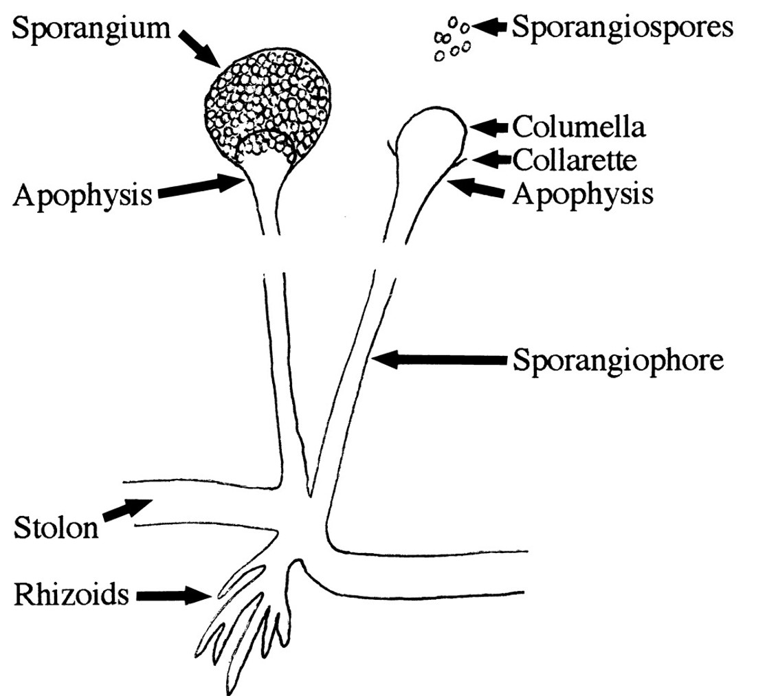
a)Sporangiospores
Sporangiospores are single celled spores produced inside the special sac-like structures called Sporangium (Sporangia Pl.). Sporangia are produced at the tip of special hyphae called Sporangiophores. A sterile dome-like structure at the tip of a sporangiophore or within a sporangium is called Columella. A funnel-shaped swelling of a sporangiophore, immediately below the columella, can be seen in some fungi and are called Apophysis.
Sporangiospores can be motile or nonmotile. Non-motile sporangiospores are called Aplanospores. Motile ones are called Zoospores and their motility is due to the presence of flagella.
b)Conidiospores or conidia (Conidium)
Conidia are exogenous spores formed at the tip or side of hyphae. They can exist in two forms such as Microconidia and Macroconodia. Microconidia are small single celled conidia whereas Macroconidia are large multi celled spores.

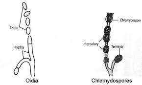
C & d)Oidia/ Oidium (Arthrospores/Thallospores) and Chlamydospores
Both Oidia and Chlamydospores are produced by the disjoining or fragmentation of vegetative hyphae. In oidia formation, hyphal cells simply disjoin from the apical regions and each fragmented cell acts as spore, giving rise to new fungi. In the case of Chlamydospores, cells become enveloped by a thick wall before hyphal fragmentation. These thick walled, single celled Chlamydospores are highly resistant to the adverse conditions and can be terminal or intercalary.
e) Blastospores
Blastospores are spores formed by budding. It is also known as a blastoconidium (pl. blastoconidia). An example of a fungus that forms blastospores is Candida albicans.
Other means of asexual reproduction in fungi include formation of Sclerotia and Rhizomorphs. These asexual methods are usually used to overcome unfavourable environmental conditions.
The sclerotia are resistant and perennating bodies. Each sclerotium is a cushion-like structure of compact mycelium. They survive for many years. They give rise to new mycelia on the approach of favourable conditions.
Rhizomorphs are rope-like modified mycelium, also resistant to unfavourable conditions and give rise to new mycelia even after several years on the approach of favourable conditions.
Youtube Link: https://www.youtube.com/watch?v=yX7Ba5VCI9w


